The human eye
The human eye
The eye is one of our most important sense organs. When stimulated by light, electric impulses are produced by its receptors.
Grades 6 – 12
mozaLink
/Weblink
Scenes
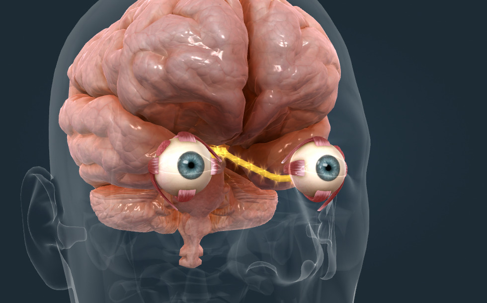
The mechanism of vision
- pupil - It is the eye’s aperture, and the iris acts like a shutter that controls the amount of light falling on the retina. In strong light, the pupil is contracted by the smooth muscles of the iris, while in low light, it is dilated. The pupillary light reflex is an unconditioned reflex, its centre is located in the brain stem. Abnormal operation of the pupillary reflex, therefore, indicates the injury of the brain stem.
- optical centre - The centre is located in the cortex of the occipital lobe.
- optic nerve - Also known as Cranial nerve II. It transmits impulses produced in the receptors of the retina to the brain.
- optic chiasm - It is the part of the brain where the optic nerves partially cross. Impulses from the inner (nasal) sides of each retina cross over to the opposite side of the brain. Impulses from the external (temporal) side, on the other hand, stay on the same side.
- extraocular muscles - Striated muscles that move the eyeballs.
- lacrimal gland - It produces tears, which serves to moisten and clean the eyes, and it plays an important role in certain emotional reactions.
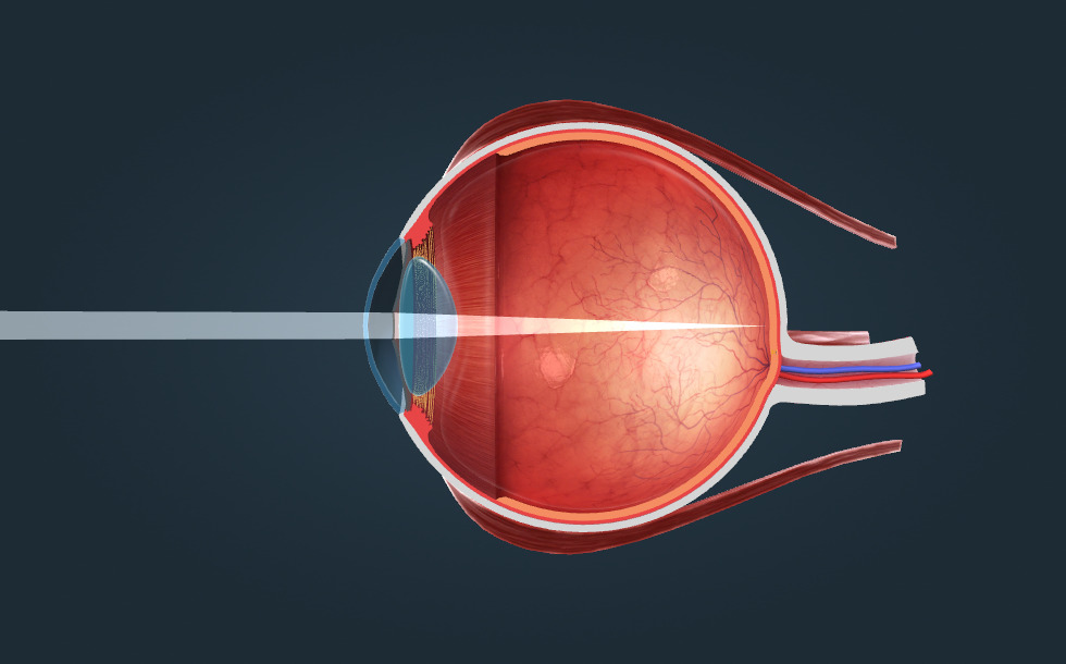
Cutaway
- iris - It is the continuation of the choroid. Its smooth muscle ensures the adaptation to changing levels of light: in strong light, the pupil contracts, while in weak light, it dilates. The iris contains pigments which give the human eyes their individual colour.
- pupil - It is the eye’s aperture, and the iris acts like a shutter that controls the amount of light falling on the retina. In strong light, the pupil is contracted by the smooth muscles of the iris, while in low light, it is dilated. The pupillary light reflex is an unconditioned reflex, its centre is located in the brain stem. Abnormal operation of the pupillary reflex, therefore, indicates the injury of the brain stem.
- lens - It is a convex lens with variable focal distance. Its flexibility allows it to become more curved when looking at a nearby object, while it can be flattened by the ciliary zonules. This provides a sharp image on the retina. As we age, the lens loses its flexibility, thus, looking at nearby objects (which requires the lens to become more curved) puts a strain on the eyes in old age. This is called presbyopia. A cataract is a grey clouding that develops in the lenses, making it opaque, which may lead to blindness.
- ciliary zonules - They hold the lens and follow the movements of the ciliary muscles. When looking at a nearby object, the ciliary muscles contract, ciliary zonules therefore become less tense and the lens becomes more curved. When looking at a distant object, the ciliary muscles relax, ciliary zonules become tense and flatten the lens.
- ciliary body - It is the continuation of the choroid. Its smooth muscles (ciliary muscles) ensure the accommodation of the eye lens to the distance of the object viewed. When looking at a nearby object, the ciliary muscles contract, ciliary zonules therefore become less tense and the lens becomes more curved. When looking at a distant object, the ciliary muscles relax, ciliary zonules become tense and flatten the lens. That is, the muscles of the ciliary body work when looking at nearby objects, and this can cause eye fatigue. It helps to relax the ciliary muscles to focus on a distant object intermittently.
- cornea - The continuation of the sclera. It is a transparent layer, where light entering the eye is refracted by the greatest angle.
- anterior chamber - It contains aqueous humour. When there is too much of this liquid, ocular hypertension occurs which causes glaucoma. This in turn, may lead to blindness as hypertension can destroy the retina.
- vitreous chamber - A chamber filled with a transparent gel called vitreous humour. Light reaches the retina through this chamber.
- macula lutea - The area of the retina responsible for visual acuity. The inverted miniature image of objects is formed here. In the centre of the macula, there are only cone cells, towards the edge the number of rod cells increases.
- blind spot (scotoma) - This is the point where the optic nerve passes through the retina, consequently, a small part of the image formed on the retina is missing (this is why it is known as the 'blind spot'). However, the brain 'fills in' this gap so we perceive a complete image.
- sclera - A very durable layer, its continuation at the front of the eye is the cornea.
- choroid - A layer containing blood vessels supplying the eye. Its continuation at the front of the eye is the ciliary body and the iris.
- retina - It contains receptors called cone cells and rod cells. The area of the retina responsible for visual acuity is called the macula lutea. The blind spot is the place where the optic nerve passes through the retina, it contains neither cone cells nor rod cells.
- optic nerve - Also known as Cranial nerve II. It transmits impulses produced in the receptors of the retina to the brain.
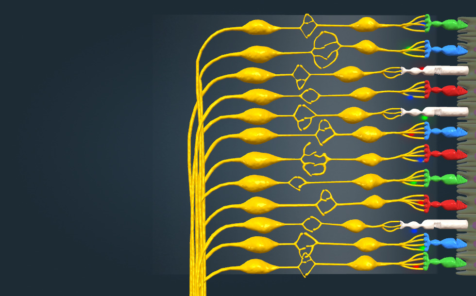
Retina
- cone cell - They contain three types of light-sensitive pigment, making them sensitive for red, green or blue light. Their stimulation threshold is higher than that of rods, this is why we perceive colours much less bright at dusk. In the centre of the macula, there are only cone cells, towards the edge the number of rod cells increases.
- rod cell - These cells cannot differentiate between colours, as they are stimulated by light of all wavelengths. Their stimulation threshold is much lower than that of cone cells: they even respond to a single photon. Therefore they are also active when there is not enough light for cone cells. In the centre of the macula, there are only cone cells, towards the edge the number of rod cells increases.
- bipolar cell - They transmit the impulses of the receptors to the ganglion cells.
- ganglion cell - It is stimulated by bipolar cells, its axon form the optic nerve.
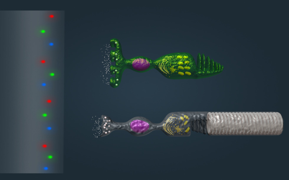
Receptors
- green-sensitive cone cell
- rod cell - These cells cannot differentiate between colours, as they are stimulated by light of all wavelengths. Their stimulation threshold is much lower than that of cone cells: they even respond to a single photon. Therefore they are also active when there is not enough light for cone cells. In the centre of the macula, there are only cone cells, towards the edge the number of rod cells increases.
- membrane discs - They are lined with a large amount of rhodopsin, which consists of a protein called ‘opsin’ and a photosensitive derivative of Vitamin A called ‘retinal’. Light causes cis retinal to become trans-retinal, which induces a signal transmission process in the cell. The cell becomes hyperpolarised, resulting in the release of a temporarily decreased amount of neurotransmitters (glutamate).
- invagination - They are lined with a large amount of iodopsin, which is similar to the rhodopsin found in rod cells. It differs only in the protein component – it has three versions with different protein components sensitive for green, red or blue light. Rhodopsin and iodopsin also contain a photosensitive derivative of Vitamin A called ‘retinal’. Light causes cis retinal to become trans retinal, which induces a signal transmission process in the cell. The cell becomes hyperpolarised, resulting in the release of a temporarily decreased amount of neurotransmitters (glutamate).
- mitochondrion - It is responsible for the cells’energy supply, it produces ATP.
- nucleus - It contains the genetic material of the cell, which controls the cell’s metabolic processes.
- synaptic vesicles - They contain neurotransmitters called ‘glutamate’, which block bipolar cells. In darkness glutamate is released continuously. Light causes the receptor to hyperpolarise and release less glutamate. Therefore the bipolar cell is unblocked and it produces an impulse.

Eyes
- eyelid - They are covered with thin skin on the outside and conjunctive tissue inside. Their role is to provide mechanical protection for the eyeballs, to keep them warm and prevent them from drying out.
Visible light is electromagnetic radiation that has a wavelength within a range of about 380 to 800 nm. Light of 380 nm in wavelength is seen as violet, while light of 800 nm in wavelength is seen as red. Light is perceived by the eyes. The impulses caused by light in the eyes are transmitted to the brain by the optic nerve, also called Cranial nerve II. The optic nerves are partially crossed in the optic chiasma, therefore, impulses from the inner sides of each retina cross over to the opposite side of the brain. Impulses from the external side, on the other hand, stay on the same side. After entering the brain, optic nerve fibres run to the visual cortex found in the occipital lobe via the optic tract. The sense of light is formed in the cerebral cortex.
The amount of light entering the eyes is regulated by the pupillary light reflex. In strong light, the pupil is contracted by the smooth muscles of the iris, while in low light, it is dilated. The pupillary light reflex is an unconditioned reflex, its centre is located in the brain stem. Abnormal operation of the pupillary reflex therefore is an indication of injury to the brain stem. The eyeballs are moved by the extraocular muscles. These are striated muscles under voluntary control.
The vitreous chamber forms the main mass of the eye. The cross-section of the eye shows three main layers. The outermost is the sclera, a very durable layer of connective tissue, which continues in the transparent cornea. This is where light entering the eye is refracted by the greatest angle.
The second layer is the choroid, which contains blood vessels that supply the eye. Its continuation at the front of the eye is the ciliary body and the iris. The smooth muscles of the iris are responsible for the pupillary light reflex. The iris contains pigments which lend human eyes their colour.
The smooth muscles of the ciliary body ensure the accommodation of the eye lens to the distance of the object viewed by changing its curvature.
The lens is connected to the ciliary body through the ciliary zonules. The ciliary body is also responsible for producing the aqueous humour, the liquid that fills the anterior chamber. If the drainage of the aqueous humour is insufficient, the pressure increases in the eye, which causes glaucoma. In serious cases, it may lead to blindness.
The innermost layer is the retina. This is where an inverted miniature image is formed of the object viewed; this is created by the lens. Its receptors are called rod cells and cone cells. The area of the retina responsible for visual acuity is called the macula lutea: in its centre there are only cone cells, while around the edge there are more rod cells. The blind spot is the place where the optic nerve passes through the retina. There are no receptor cells here. The impulses produced by the receptors in the retina are transmitted to the brain by the nerve fibres in the optic nerve.
The receptors in the retina are called rod cells and cone cells. They transmit impulses to the bipolar cells, which stimulate ganglion cells. The axons of the ganglion cells form the optic nerve.
The light-sensitive pigment in rod cells is rhodopsin, which consists of a protein called ‘opsin’ and a Vitamin A derivative called ‘retinal’. Rhodopsin is sensitive to light of any wavelength, therefore rod cells cannot differentiate between colours.
The stimulation threshold of rod cells is low, a single photon is enough to stimulate them, thus they work in weak light.
The three types of cone cells are sensitive to red, green or blue light. Their photosensitive pigment is iodopsin, which is similar to rhodopsin but contains a different protein. The stimulation threshold of cone cells is higher than that of rod cells; they are not active in low light. This is why we lose colour vision at dusk.
We can see dim stars better using our peripheral vision, because this way their images are not formed on the macula, but on areas richer in sensitive rod cells. Colour blindness means that a type of cone cell is missing or does not work properly. The most common type of colour blindness is red-green dichromacy, difficulty in distinguishing between red and green colours. When all three types of cones are involved, total colour blindness or monochromacy occurs.
In darkness, cones and rods continuously release a neurotransmitter called glutamate, which blocks bipolar cells. Light causes the receptor cells to hyperpolarise, that is to produce an electric impulse. This stops the release of glutamate, therefore bipolar cells are unblocked and they produce action potentials.

