Your cart is empty
The ear and the mechanism of hearing

The ear and the mechanism of hearing
The ear converts the vibrations of air into electric signals which are then processed by the brain.
Grades 6 – 12
mozaLink
/Weblink
Questions
- True or false? The cochlea is filled with a fluid which is vibrated by the movement of the stirrup.
- True or false? Sounds of the same frequency are always absorbed at the same place in the cochlea.
- True or false? The outermost ossicle, connected to the eardrum, is the anvil.
- True or false? Sound waves generate signals in the Eustachian tube.
- True or false? The Eustachian tube is also known as the auditory tube.
- True or false? The base of the stirrup fits into the oval window of the cochlea.
- What frequency range can a healthy ear perceive?
- What kind of sounds are absorbed at the base of the cochlea?
- In which lobe of the cerebral cortex is the auditory cortex located?
- What connects the tympanic cavity with the pharyngeal cavity?
- What separates the external ear from the middle ear?
- In which part of the ear are electric signals generated?
- Where is the cochlea located?
- What type of tissue comprises the bulk of the auricle?
- Which of the following bones is not one of the ossicles?
- Where is the sense of sound produced?
- Where are lower frequency vibrations generated by deep sounds absorbed?
- Which cranial nerve is called the vestibulocochlear nerve?
- Where are the ossicles located?
Scenes
The mechanism of hearing
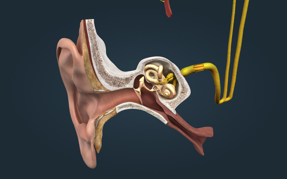
- auricle - It guides sound waves into the external auditory canal. It is composed mainly of cartilage tissue.
- external auditory canal - It channels sound waves to the eardrum. The skin lining the auditory canal produces ear wax, which protects it against injuries and infections. Excessive earwax may impede the passage of sound in the ear canal, causing temporary hearing loss.
- middle ear - It contains the tympanic cavity and the ossicles. It is connected with the pharyngeal cavity by the Eustachian tube.
- inner ear - It has a vital role in balancing and hearing.
- cochlear nerve - The 8th cranial nerve, carrying signals from the cochlea of the inner ear to the brain. This nerve also carries information responsible for balancing, therefore it is also called the vestibulocochlear nerve.
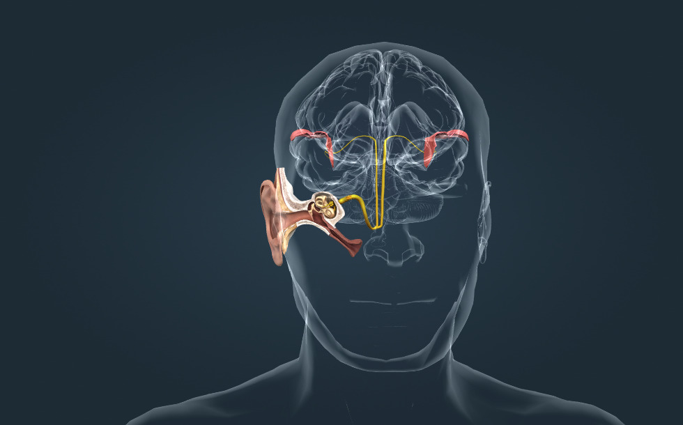
- auditory pathway - The continuation of the auditory nerve in the brain. Its axons transmit a signal to the auditory cortex through the thalamus.
- auditory cortex - A cortical region of the brain, located in the temporal lobe, it processes sound. Its pitch-sensitive areas are activated by different pitches of sound.
- Eustachian tube - It connects the nasal cavity with the middle ear (tympanic cavity) and allows the pressure to equalize between the middle ear and the outside world. It usually opens during swallowing; when it is permanently closed, air pressure in the middle ear drops, making the ear feel blocked. When external air pressure changes we can hear a popping sound: the Eustachian tube opens and air flows in the tympanic cavity (if the external air pressure is higher) or out of it (if the external air pressure is lower).
Ear
- auricle - It guides sound waves into the external auditory canal. It is composed mainly of cartilage tissue.
- external auditory canal - It channels sound waves to the eardrum. The skin lining the auditory canal produces ear wax, which protects it against injuries and infections. Excessive earwax may impede the passage of sound in the ear canal, causing temporary hearing loss.
- middle ear - It contains the tympanic cavity and the ossicles. It is connected with the pharyngeal cavity by the Eustachian tube.
- inner ear - It has a vital role in balancing and hearing.
- cochlear nerve - The 8th cranial nerve, carrying signals from the cochlea of the inner ear to the brain. This nerve also carries information responsible for balancing, therefore it is also called the vestibulocochlear nerve.
- auditory pathway - The continuation of the auditory nerve in the brain. Its axons transmit a signal to the auditory cortex through the thalamus.
- auditory cortex - A cortical region of the brain, located in the temporal lobe, it processes sound. Its pitch-sensitive areas are activated by different pitches of sound.
- Eustachian tube - It connects the nasal cavity with the middle ear (tympanic cavity) and allows the pressure to equalize between the middle ear and the outside world. It usually opens during swallowing; when it is permanently closed, air pressure in the middle ear drops, making the ear feel blocked. When external air pressure changes we can hear a popping sound: the Eustachian tube opens and air flows in the tympanic cavity (if the external air pressure is higher) or out of it (if the external air pressure is lower).
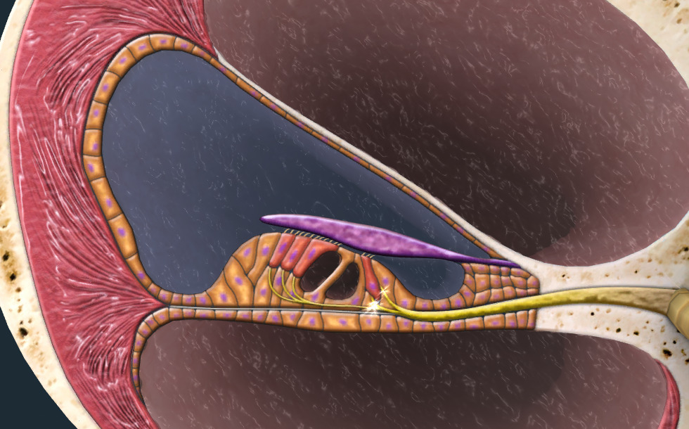
Ossicles
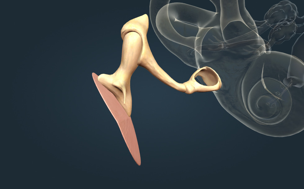
- eardrum - A membrane that separates the external ear from the middle ear. Sound waves make it vibrate, this vibration is transmitted to the ossicles. When a myringotomy is performed, a small incision is made on the eardrum, allowing the drainage of pus from the inflamed middle ear.
- hammer (malleus) - The outermost of the ossicles, it transmits the vibration of the eardrum to the anvil.
- anvil (incus) - The central ossicle, it transmits the vibration of the hammer to the stirrup.
- stirrup (stapes) - The innermost ear bone, it transmits the vibration of the anvil to the cochlea. It is the smallest bone of the human body.
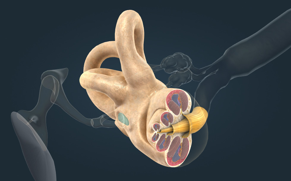
Cochlea
- 3 semicircular canals - They detect angular acceleration of the head. When the head turns to any direction, a signal is created in the receptors of the semicircular canals, this signal is then transmitted to the brain by the axons of the cochlear nerve (vestibulocochlear nerve).
- scala vestibuli - The fluid in the scala vestibuli (the perilymph) is vibrated by the stirrup. The vibration of the fluid spreads towards the tip of the cochlea.
- scala media - It is separated from the scala vestibuli by the Reissner´s membrane, and from the scala tympani by the basilar membrane. It is filled with fluid (endolymph).
- scala tympani - It is filled with fluid (the perilymph). Vibration spreads from the tip towards the base of the cochlea in this canal.
- cochlear nerve - The 8th cranial nerve, carrying signals from the cochlea of the inner ear to the brain. This nerve also carries information responsible for balancing, therefore it is also called the vestibulocochlear nerve.
- round window - It is covered with a connective tissue membrane. Vibration spreads in the liquid filling the lower cochlear canal towards the round window. The round window is the ´exit´ of the cochlea.
- oval window - It is covered with a connective tissue membrane called the oval membrane. The base of the stirrup fits in snugly within it; the vibration of the stirrup is transmitted to the liquid filling the upper cochlear canal by the membrane. The oval window is the ´entrance´ of the cochlea.
Organ of Corti
- hair cell - When vibration is absorbed, the basilar membrane and the tectorial membrane move in relation to each other. The tectorial membrane is pushed against the hair cells in the Organ of Corti and bends them, which generates a signal in the cells. Continuous exposure to noise may cause the destruction of hair cells leading to permanent hearing loss. This is why adequate workplace noise protection is important.
- tectorial membrane - When vibration is absorbed, the basilar membrane and the tectorial membrane move in relation to each other. The tectorial membrane is pushed against the hair cells in the Organ of Corti and bends them which generates a signal in the cells.
- basilar membrane - It absorbs vibration spreading in the cochlear liquid and it starts to vibrate. It causes the basilar membrane and the tectorial membrane move in relation to each other.
- nerve fibres
Tonotopy
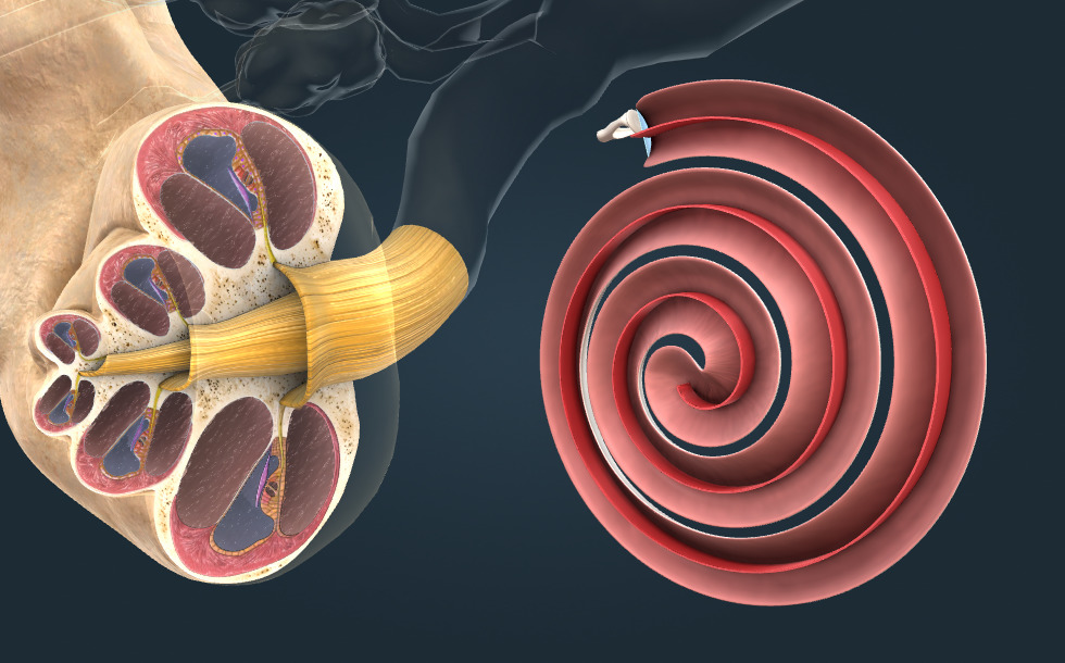
According to tonotopy, the pitch is determined by where the vibration is generated along the basilar membrane in the inner ear. Higher frequency sounds cause vibrations of higher frequency in the liquid, which are absorbed in the initial section of the membrane. Lower frequency vibrations generated by deep sounds enter the cochlea and are absorbed closer to the tip. When a vibration is absorbed, an electrical signal is produced, which is transmitted into the brain. The pitch of the sound is encoded by the site of absorption.
Georg von Békésy (1899–1972), a Hungarian-American biophysicist, proved this theory through various experiments. His discoveries significantly advanced the understanding of the mechanism of hearing. In 1961, he was awarded the Nobel Prize in Physiology or Medicine 'for his discoveries of the physical mechanism of stimulation within the cochlea.'
Narration
Sound is the vibration of air perceived by our ears. Healthy ears can perceive sound waves of frequencies from about 20 to 20,000 Hz. This range will become narrower due to ageing or noise exposure.
Sound waves create signals in the inner ear, which are transmitted to the auditory cortex by the cochlear nerve and auditory pathway. The sense of sound is produced in the auditory cortex.
Sound waves are directed into the external auditory canal by the auricle. Sound waves cause the eardrum, which closes the auditory canal, to vibrate. The vibration of the eardrum is transmitted to the cochlea by the ossicles - the hammer, the anvil and the stirrup.
The base of the stirrup fits snugly into the oval window of the cochlea. The basilar membrane is located inside the cochlea. It runs along the tip of the cochlea, where it turns back and continues in Reissner's membrane. The membranes divide the cochlea longitudinally into three cavities: the scala tympani, the scala media and the scala vestibuli.
The chochlea is filled with a fluid, which is vibrated by the stirrup. Higher frequency sounds cause vibrations of higher frequency in the liquid, which are absorbed in the initial section of the membrane. Lower frequency vibrations generated by deep sounds enter the cochlea and become absorbed closer to the tip. When a vibration is absorbed, an electrical signal is produced which is transmitted into the brain. The pitch of the sound is encoded by the site of absorption: this is called tonotopy.
Electrical signals are generated in the organ of Corti. Vibrations spreading inside the cochlea push the tectorial membrane against the hair cells found on the basilar membrane and bend them, generating a signal in the cells. Thus the organ of Corti transforms vibrations into electrical signals, which are transmitted into the brain by the cochlear nerve, and then into the auditory cortex by the auditory pathway. Finally, the sense of sound is produced in the cerebral cortex.